Back Of Neck Anatomy Glands - Hodgkin Lymphoma Facts Seattle Cancer Care Alliance : Submandibular triangle carotid and muscular triangles sternocleidomastoid region.
Back Of Neck Anatomy Glands - Hodgkin Lymphoma Facts Seattle Cancer Care Alliance : Submandibular triangle carotid and muscular triangles sternocleidomastoid region.. And then also, you've got these vessels, which obviously run in the anterior triangle. In the front, the neck extends from the bottom part of the mandible (lower jaw bone) to the salivary glands the submandibular salivary glands and the tail of the parotid salivary gland are located in the upper part of the neck. This atlas of otolaryngologic anatomy on an mri of the face and neck was. The neck is the area between the skull base and the clavicles. Trachea and thyroid gland and also form the anterior boundaries of the neck levels.
In some cases, inflammation of neck glands may occur due to hodgkin's. Clinically, surface anatomy is used to split the neck into anterior and posterior triangles which provide clues as to the location of specific structures. The endocrine system includes all of the glands of the body and the hormones produced by those glands. Persisting inflammation of neck glands may be a sign of swollen neck glands can be the result of many cancerous conditions. The vocal cords are attached to the back of this prominence, and muscles attached to the oblique line, on the outer surface of the cartilage, to the.
Lymph Nodes Healthdirect from media.healthdirect.org.au Anatomy and function neck, regions of the lower face, cervical spine, head joints,.during muscle traction, the cheeks are pulled together, which makes food move back and forth between the.the parotid gland, which is one of the 3 major salivary glands. It includes the infrahyoid musculature and thyroid glands with the parathyroid glands. Anatomy of the human body. And what's the deal with the retropharyngeal space? Click now to study the muscles, glands and organs of the neck at kenhub! Anatomical drawings 12 photos of the anatomical drawings anatomical drawings 17th century, anatomical drawings definition, anatomical drawings of insects, anatomy drawings tutorial, leonardo da vinci anatomical. The deep muscles of the back and the suboccipital muscles are supplied by the posterior primary rami of the spinal nerves. All anatomical structures of the terminologia anatomica are translated in french, english, spanish, japanese, portuguese, polish, russian, german, italian and chinese.
Head and neck anatomy is important when considering pathology affecting the same area.
This module can be used as a medical dictionary. This article describes the anatomy of the head and neck of the human body, including the brain, bones, muscles, blood vessels, nerves, glands, nose, mouth, teeth, tongue, and throat. 3.6 ) and 120° in the female ( fig. There are lymph nodes on the back of the neck which may become inflamed with infections both viral and bacterial. Want to learn more about it? This atlas of otolaryngologic anatomy on an mri of the face and neck was. Anatomical observation and palpation sk… surface anatomy: Major glands are the primary glands providing the oral cavity and its structure moistening, lubrication, and protection. Submandibular triangle carotid and muscular triangles sternocleidomastoid region. 803 x 1024 jpeg 192 кб. The lymphatics of the head, face, and neck. Such a division has an anatomical substrate, because the deep fascia of the neck sends in frontal 8. The vocal cords are attached to the back of this prominence, and muscles attached to the oblique line, on the outer surface of the cartilage, to the.
The vocal cords are attached to the back of this prominence, and muscles attached to the oblique line, on the outer surface of the cartilage, to the. What are the pretracheal and prevertebral fascia? Learning the anatomy of the neck is a usually the thyroid gland consists of right and left lateral lobes which are joined across the midline by the normal thyroid gland is occasionally visible and, although it has a soft consistency, it can. The parotid gland locates anterior to the outer ear, the submandibular gland is located below the oral. The lymphatics of the head, face, and neck.
Overview Of The Head And Neck Region Amboss from media-us.amboss.com Frontal view of the muscles and glands of the human neck. Neck, in land vertebrates, the portion of the body joining the head to the shoulders and chest. Learning the anatomy of the neck is a usually the thyroid gland consists of right and left lateral lobes which are joined across the midline by the normal thyroid gland is occasionally visible and, although it has a soft consistency, it can. Anatomy and function neck, regions of the lower face, cervical spine, head joints,.during muscle traction, the cheeks are pulled together, which makes food move back and forth between the.the parotid gland, which is one of the 3 major salivary glands. It runs down the back part of the neck, and opens into the external jugular vein just below the middle of its course. The deep muscles of the back and the suboccipital muscles are supplied by the posterior primary rami of the spinal nerves. Clinically, surface anatomy is used to split the neck into anterior and posterior triangles which provide clues as to the location of specific structures. And then also, you've got these vessels, which obviously run in the anterior triangle.
Anatomy of the human body.
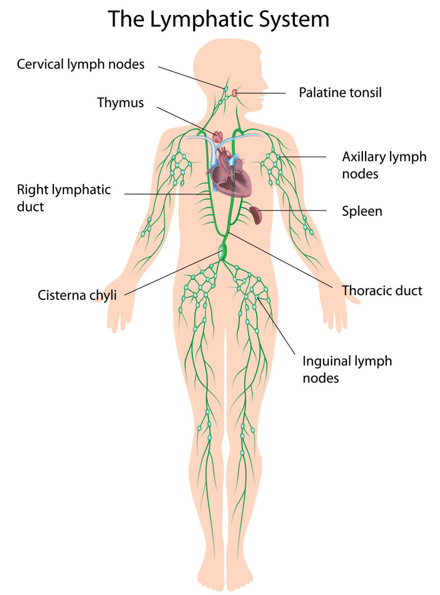
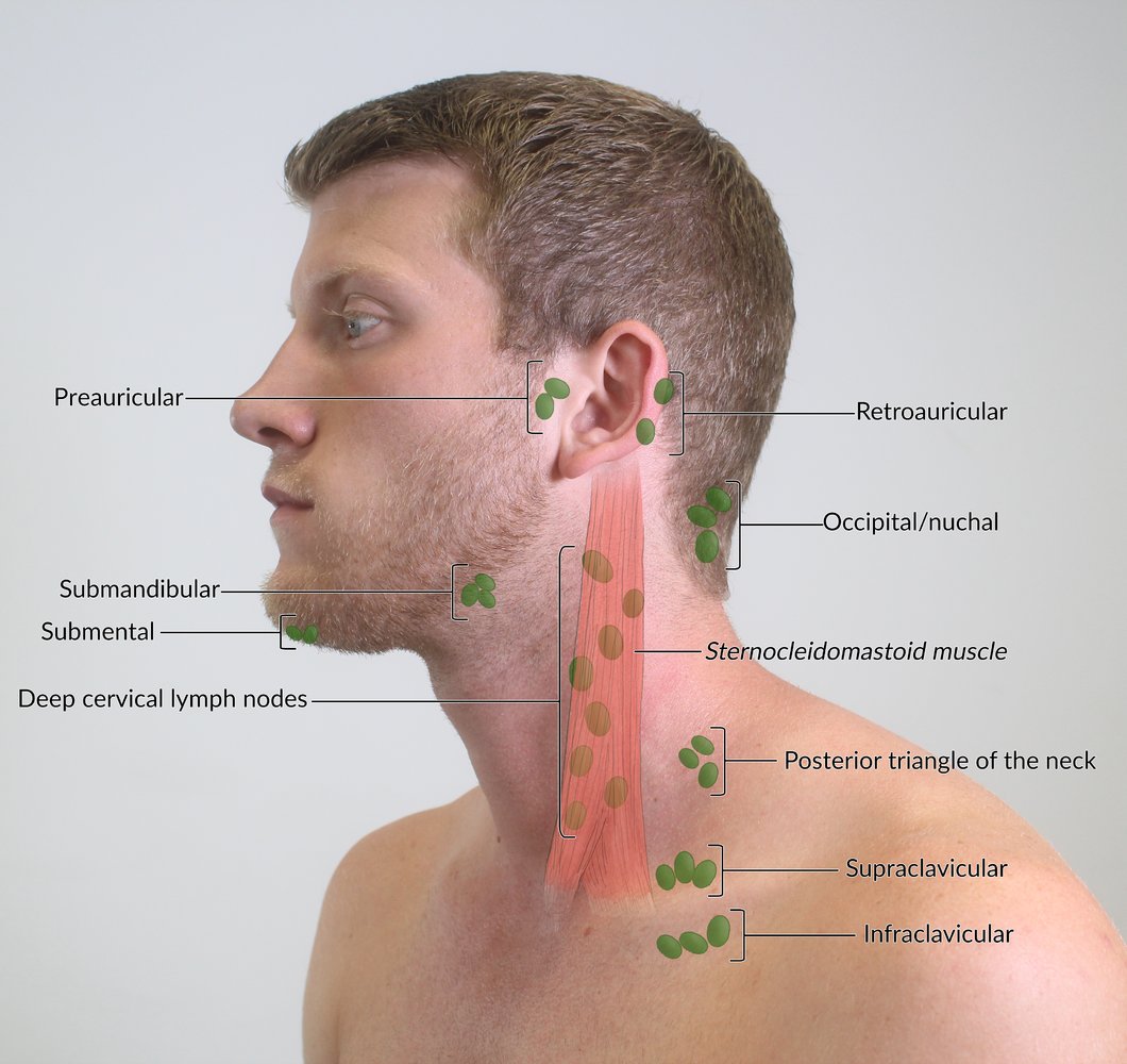
Living anatomy of the anterior and lateral aspects of the neck. We've also got the parathyroid glands behind the thyroid. Head and neck anatomy is important when considering pathology affecting the same area. And then also, you've got these vessels, which obviously run in the anterior triangle. Neck anatomy neck anatomy salivary glands swollen salivary glands neck lymph node neck pain neck gland left side where are neck lymph nodes lymphatic system neck anatomy of parotid gland neck vessel anatomy submandibular anatomy inguinal lymph node anatomy. In the front, the neck extends from the bottom part of the mandible (lower jaw bone) to the salivary glands the submandibular salivary glands and the tail of the parotid salivary gland are located in the upper part of the neck. All anatomical structures of the terminologia anatomica are translated in french, english, spanish, japanese, portuguese, polish, russian, german, italian and chinese. The occipital glands (lymphoglandulæ occipitales), one to three in nu ber, are placed on the back of the head close to the margin of the trapezius and resting on the insertion of the semispinalis capitis. The deep muscles of the back and the suboccipital muscles are supplied by the posterior primary rami of the spinal nerves. Via the retromandibular vein and the. The lymphatics of the head, face, and neck. There are lymph nodes on the back of the neck which may become inflamed with infections both viral and bacterial. Such a division has an anatomical substrate, because the deep fascia of the neck sends in frontal 8.
Frontal view of the muscles and glands of the human neck. How is it organised in the neck? The anatomy of the head and neck is complex because so many different functional structures are located close to each other. Some important structures contained in or passing through the neck include the seven cervical vertebrae and enclosed spinal cord, the jugular veins and carotid arteries, part of the esophagus, the larynx. 3.6 ) and 120° in the female ( fig.
Lymphatic Drainage Of The Head And Neck Ppt Video Online Download from slideplayer.com Major glands are the primary glands providing the oral cavity and its structure moistening, lubrication, and protection. The anterior jugular vein (v. This article describes the anatomy of the head and neck of the human body, including the brain, bones, muscles, blood vessels, nerves, glands, nose, mouth, teeth, tongue, and throat. The vocal cords are attached to the back of this prominence, and muscles attached to the oblique line, on the outer surface of the cartilage, to the. Anatomy and function neck, regions of the lower face, cervical spine, head joints,.during muscle traction, the cheeks are pulled together, which makes food move back and forth between the.the parotid gland, which is one of the 3 major salivary glands. Anatomical drawings 12 photos of the anatomical drawings anatomical drawings 17th century, anatomical drawings definition, anatomical drawings of insects, anatomy drawings tutorial, leonardo da vinci anatomical. The neck is a complex anatomic region between the head and the body. Anatomy of the human body.
The anterior jugular vein (v.
Cervical fascia and interfascial spaces in the neck. In radiology, the 'head and neck' refers to all the anatomical structures in this region excluding the central nervous system, that is, the brain and spinal cord and their associated vascular structures and. The lymphatics of the head, face, and neck. The endocrine system includes all of the glands of the body and the hormones produced by those glands. The major salivary glands are the parotid (accounting for 90% of all salivary gland tumors), submandibular or submaxillary (about 10%), and sublingual glands (1%). Choose from 500 different sets of flashcards about neck anatomy back neck upper on quizlet. The head rests on the top part of the vertebral column, with the skull joining at c1. It includes the infrahyoid musculature and thyroid glands with the parathyroid glands. Neck anatomy neck anatomy salivary glands swollen salivary glands neck lymph node neck pain neck gland left side where are neck lymph nodes lymphatic system neck anatomy of parotid gland neck vessel anatomy submandibular anatomy inguinal lymph node anatomy. There are lymph nodes on the back of the neck which may become inflamed with infections both viral and bacterial. And then also, you've got these vessels, which obviously run in the anterior triangle. This atlas of otolaryngologic anatomy on an mri of the face and neck was. How is it organised in the neck?


Posting Komentar
0 Komentar